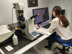
The HPI LMU was recently upgraded with the purchase of an inverted confocal microscope, which was acquired through the Stavros Niarchos foundation.
Technical specifications of the Leica TCS-SP8 inverted Confocal Microscope
The Leica TCS-SP8 confocal system is appropriate for imaging of fixed samples and live cell imaging. In the near future the system will be upgraded with a tandem scanner ideal for fast live cell imaging, an environmental chamber with controlled temperature, humidity and CO2 concentration, and a navigator for imaging unlimited number of positions for multi-well projects. The upgrading is funded by the Bioimaging-GR program. More specifically, the technical characteristics of the system are:
- Automated scanning stage for acquisition of optical sections in Z axis
- Automated and coded disc for 6 objective lenses
- Flat apochromatic lenses of excellent quality, corrected to infinite and to UV. Dry lens: 10X (NA 0.4), Oil-immersion lenses: 40X (NA 1.3) and 63X (NA 1.4).
- Head, equipped with flat ocular lenses 10X
- Halogen light source 12V, 100W for transmitted light applications.
- External Hg light source 120W for fluorescence applications, equipped with diaphragms for volume fluctuations and optical fibers for light transfer.
The confocal scanning system contains:
- Scanning head for for scanning with high analysis at a) xyz-axis, b) xzy-axis, c) temporal image scanning at xt, xyt, and xyzt, d) zoom between 1x and 64X and e) spectral scanning xλ, xyλ and xzλ. Image resolution up to 8192 x 8192 pixels, scan speed up to 1800 Hz
- Solid state laser, lines 405, 488, 514, 522, 638 nm
- Two photo-multipliers (PMTs) for confocal detection, a HyD3 detector of enhanced sensitivity and quantosome yield, and a PMT trans detector for phase-contrast detection. All these detectors have independent gain, offset and spectral position adjustments.
- The system’s software LAS X allows the full control of the confocal microscope, laser sources and scanning head, image acquisition and storage, 3D image reconstruction from serial optic sections, creation of time- lapse movies and image processing.